HT-HUD Device Split View

HT-HUD: Advanced Skin Analysis through Thermal Imaging

A revolutionary method for the early detection of skin cancer.

Sweden has one of the highest incidences of skin cancer in the world, with a continuously increasing trend. Only Australia and New Zealand have higher figures. Annually, approximately 60,000 cases are diagnosed in Sweden, with 500 fatalities. UV radiation from the sun is the primary risk factor, and despite increased awareness of sun protection, the number of cases continues to rise.

Technical Components

IR Camera

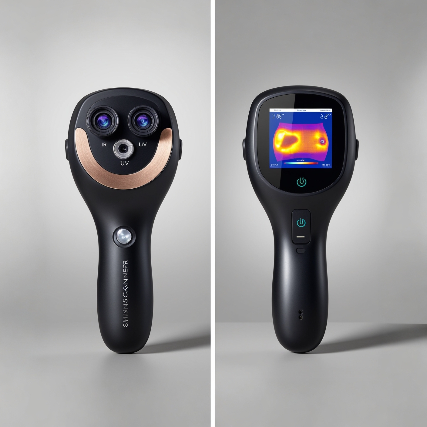
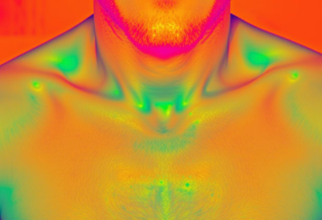
Detects infrared radiation from the skin, providing a detailed image of the skin's temperature distribution. Measures temperature differences with high precision to detect anomalies.

UV Camera

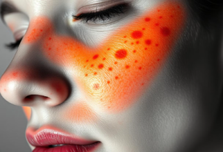
Displays the skin's reaction to ultraviolet light, revealing hidden changes not visible to the naked eye. Particularly effective in identifying early skin changes.

Visible Light Camera

Provides a standard visual image of the skin's surface, complementing the information from IR and UV cameras. Used for documentation and long-term follow-up.

Supporting Systems

1

AI Integration

Advanced artificial intelligence algorithms analyze images and data to assist in diagnosis.

2

Database Connection

Secure storage and access to patient data and examination results.

3

Global Reach

Connected to healthcare providers worldwide for comprehensive care.

Heating System and Measurement

Infrared Heating Device/IPL

  • Utilizes two different techniques:
    • Laser: A single type of concentrated light for high precision
    • IPL (Intense Pulsed Light): Multiple types of light for broader treatment
  • Controlled heating of the skin area
  • Enables analysis of heat distribution

Infrared Thermometer

  • Measures skin temperature with high precision
  • Detects small temperature differences
  • Monitors both heating and cooling phases
  • Vital for identifying abnormal thermal patterns

Integrated Display

  • Shows real-time data from all sensors
  • Displays temperature measurements
  • Provides an intuitive interface for healthcare professionals

Examination Process with HT-HUD

1

Patient Registration

  • Patient presents ID.
  • Registration in the system's database.
  • Personal data securely stored.
2

Initial Photography

  • Photography without heating.
  • Images captured using IR, UV, and standard cameras.
  • Documentation of the skin area.
3

Heating Phase

  • Controlled heating with IR or IPL.
  • Precise temperature measurement.
  • Monitoring of skin response.
4

Cooling Phase

  • Natural cooling or application of cooling pads/spray.
  • Continuous temperature monitoring.
5

Analysis

  • Comparison of pre- and post-images.
  • AI-assisted analysis.
  • Identification of anomalies.

Scientific Background

Research Basis

Based on established and recognized research:

  • Laser Doppler technology for blood flow analysis.
  • Studies on hypodermic responses and vascular changes.
  • Thermal imaging used for physiological evaluation.

Research References:

  • Roustii M et al. Trends Pharmacol Sci 2013; 34:373–84
  • Bergengren et al. Ophthalmic Plast Reconstr Surg. 2019

Scientific Principles

  • Angiogenesis affects the skin’s thermal distribution patterns.
  • Tissue density and vascularity vary in cancerous vs. healthy areas.
  • Thermal signatures change according to metabolic activity.

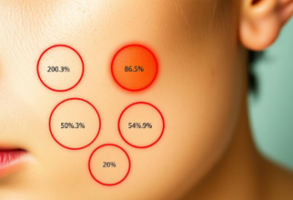
These parameters provide a measurable scientific foundation for differentiating normal and abnormal tissue behavior.

IR and UV Imaging Overview

  • Infrared (IR) and Ultraviolet (UV) techniques visualize both surface and subsurface responses.
  • IR detects heat flow and vascular function.
  • UV reveals hidden structural and pigmentation damage.

Together they create a multi-layered diagnostic perspective.

Infrared (IR) Imaging

  • Clinically used to monitor temperature variations in the skin.
  • IR is divided into three bands: IRA (near-IR), IRB (mid-IR), IRC (far-IR).
  • Each wavelength penetrates to different depths within the dermis.
  • Thermal cameras ensure safe, controlled heating at 32–35 °C.

These measurements reflect vascular activity and tissue response to thermal stimulation.

Ultraviolet (UV) Imaging

  • Highlights subsurface skin changes invisible to the naked eye.
  • Reveals collagen damage and UV-induced alterations in elasticity.
  • Maps hydration, pigmentation, and early photoaging patterns.
  • Used for before/after comparisons to show progressive damage.

UV imaging therefore plays a critical role in preventive dermatology and skin-health monitoring.

Summary

  • IR and UV imaging jointly provide objective physiological data.
  • Enable non-invasive, repeatable analysis of thermal and optical skin behavior.
  • Used in clinical, cosmetic, and research environments worldwide.

This combined methodology forms the technological foundation of HT-HUD diagnostics.

Advantages and Opportunities

Standard Photography

Standard Photography

Thermal Imaging

Thermal Imaging

UV Imaging

UV Imaging

AI-enhanced analysis

AI-enhanced analysis

Non-invasive Method

  • Painless examination.
  • No tissue damage.
  • Repeatable measurements.

Early Detection

  • Identifies changes early.
  • Enables prompt intervention.
  • Improves treatment outcomes.

AI-Assisted Analysis

  • Automated image analysis.
  • Comparison with database.
  • Continuously learning system.

Follow-up

  • Documentation over time.
  • Objective measurement data.
  • Trend analysis.

Future Outlook

Modern medical laboratory
Our vision for the future of medical diagnostics
  • 1

    Potential Development Opportunities

    HT-HUD can be further developed to include new features and improve diagnostic precision.

  • 2

    Expected Impact on Skin Cancer Diagnostics

    The system has the potential to revolutionize skin cancer diagnostics and improve patient outcomes.

  • 3

    The Role of AI Learning in Improved Diagnostics

    AI integration will improve over time by learning from new data and experiences.

Academic Validation - Lund University

Lund University Validation Letter

Scientific Validation

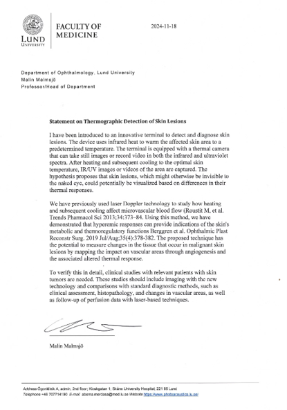
Professor Malin Malmsjö at Lund University, Department of Ophthalmology, has validated the potential of HT-HUD technology for skin cancer diagnostics. In her statement, she confirms the scientific foundation and innovative approach of the method.

Key Observations:

  • Innovative terminal for detecting skin changes.
  • Combines IR/UV imaging with thermal analysis.
  • Enables visualization of otherwise invisible skin changes.

Based on Established Laser Doppler Technology:

Research foundation: The technology is based on previous studies in:

  • Microvascular blood flow.
  • Hyperemic responses.
  • Tissue changes in malignant skin conditions.

Next Steps:

Clinical studies are planned to validate the technology through:

  • Comparison with standard diagnostics.
  • Histopathological examinations.
  • Vascular analyses.
  • Follow-up with laser technology.

"Lund University - Faculty of Medicine, 2024"

Summary

HT-HUD Device Demonstration

HT-HUD is an innovative medical device with the potential to revolutionize skin cancer diagnostics. The system offers a fast, precise, and non-invasive method for identifying potential skin changes at an early stage. By combining thermal imaging, AI integration, and database connectivity, HT-HUD provides a powerful solution to improve patient care and reduce the risk of skin cancer.

Interest Registration